The emergency department nurse assesses a 3-day-old newborn brought in by the mother. The mother states, "My baby is always so sweaty and hot and doesn't want to eat! I think something is wrong." The nurse cannot palpate a femoral pulse but notes +3 brachial pulses. Which congenital heart defect does the nurse suspect?
The emergency department nurse assesses a 3-day-old newborn brought in by the mother. The mother states, "My baby is always so sweaty and hot and doesn't want to eat! I think something is wrong." The nurse cannot palpate a femoral pulse but notes +3 brachial pulses. Which congenital heart defect does the nurse suspect?
A. Hypoplastic left heart syndrome (HLHS)
[11%]
B. Patent ductus arteriosus (PDA)
[23%]
C. Transposition of the great arteries (d-TGA)
[18%]
D. Coarctation of the aorta (COA)
[47%]
Statistics
Subject/Lesson
Subject
Child Health
Lesson
Cardiovascular
Client Need
Test Plan
Physiological Adaptation
Related Topic
Alterations in Body Systems
Item Type
Item Type
Application
Explanation
Choice D is correct. Based on the assessment findings, the infant most likely has a coarctation of the aorta. In this defect, a stricture in the aorta prevents blood flow from the left ventricle. Coarctation of the aorta usually occurs beyond the blood vessels that branch off to the upper body but before the blood vessels that lead to the lower body. Therefore, the blood flow to the upper body is abundant, whereas the flow to the lower body is diminished. This results in decreased pulses in the lower extremities, whereas bounding pulses (3+ brachial pulses) in the upper extremities.
Choice A is incorrect. Hypoplastic left heart syndrome (HLHS) is characterized by a very small, underdeveloped left atrium, ventricle, and aorta. Essentially, the entire left side of the heart is not developed. This infant will appear cyanotic and quickly show signs of heart failure but will not present with absent femoral and 3+ brachial pulses.
Choice B is incorrect. The ductus arteriosus is a normal duct in fetal circulation that allows oxygenated blood to shunt from the pulmonary artery to the aorta and bypass pulmonary circulation. The ductus arteriosus should close shortly after birth, but if it does not, it is known as a patent ductus arteriosus (PDA). These infants present with a machine-like murmur but do not have absent femoral and 3+ brachial pulses.
Choice C is incorrect. In the transposition of the great arteries (TGA), the pulmonary artery and aorta are switched, creating two separate loops for blood circulation. The deoxygenated blood enters the right atrium from the body and is sent directly back to the body via the transposed aorta. Oxygenated blood enters the left atrium from the lungs and is sent back to the lungs via the transposed pulmonary artery. These two closed loops can only be connected via an opening in the septum, either an ASD, VSD, PDA, or PFO. The child will depend on one of these openings for systemic oxygenation. The infant may present with severe cyanosis at birth but does not have absent femoral pulses and 3+ brachial pulses.
Related Videos
2 Key Topics
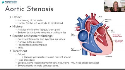
Coarctation of the Aorta (CoA)
