The nurse is caring for a client in labor. The following tracing was on the fetal heart rate monitoring strip. The nurse recognizes that this tracing is a
See the exhibit.
The nurse is caring for a client in labor. The following tracing was on the fetal heart rate monitoring strip. The nurse recognizes that this tracing is a
See the exhibit.
A. variable deceleration.
[19%]
B. late deceleration.
[53%]
C. early deceleration.
[13%]
D. normal variability pattern.
[15%]
Statistics
Subject/Lesson
Subject
Maternal & Newborn Health
Lesson
Labor/Delivery
Client Need
Test Plan
Health Promotion and Maintenance
Related Topic
Health Promotion/Disease Prevention
Item Type
Item Type
Analysis
Explanation
Choice B is correct. This strip indicates a late deceleration. Late decelerations are visually apparent and usually symmetric in shape, with a gradual decrease and return of the fetal heart rate (FHR) to baseline. Late decelerations are caused by decreased perfusion to the fetus. Maternal repositioning is an effective intervention for this nonreassuring pattern. Other interventions include oxygen administration and the administration of intravenous isotonic fluids.
Choice A is incorrect. This strip does not show a variable fetal deceleration. This strip indicates the presence of another fetal heart pattern other than variable fetal deceleration.
Choice C is incorrect. This strip does not show early fetal deceleration. This strip indicates the presence of another fetal heart pattern other than initial fetal deceleration.
Choice D is incorrect. This strip does not have a normal variability pattern. This strip indicates a non-reassuring finding.
Related Videos
2 Key Topics
Decelerations
29.1 Fetal Monitoring Basics
Additional Info
 Source : Archer ReviewSource : Archer Review
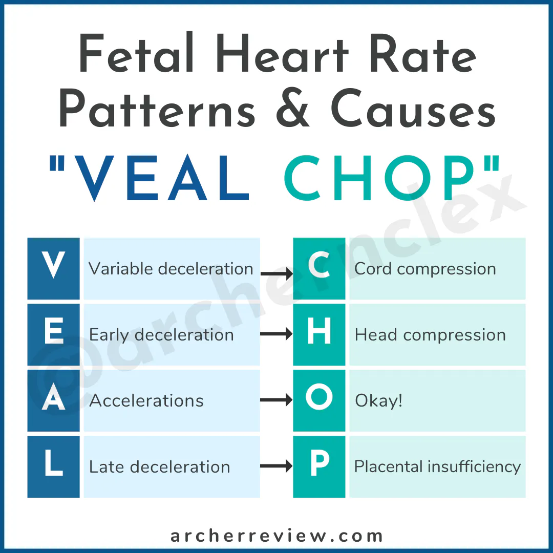
Late decelerations become more concerning when they are recurrent (present with 50% or more of the uterine contractions in a 20-minute period). Late decelerations are caused by placental insufficiency. Interventions for late decelerations include -
• Maternal repositioning (either lateral or hands and knees)
• Intravenous fluid boluses
• Administering oxygen (via nonrebreather 10 L/min)
• Discontinuing oxytocin infusion (if applicable)
